
Oral & Maxillofacial Pathology Surgery in Mumbai

What Is Oral & Maxillofacial Pathology Surgery?
Oral and maxillofacial pathology surgery involves the investigation and surgical management of abnormal tissues affecting the mouth, jaws, and surrounding facial structures. These abnormalities may include cysts, benign tumours, persistent mucosal lesions, and tissue changes that require examination to rule out precancerous conditions or cancer.
Surgery may be performed to obtain a tissue sample, remove an abnormal growth, prevent damage to adjacent structures, or establish the diagnosis. The removed tissue is examined under a microscope through histopathological analysis, which helps determine the nature of the lesion and whether additional treatment or follow-up is required.
What Conditions May Require Oral Pathology Surgery?
An oral and maxillofacial surgeon may evaluate several types of lesions and tissue abnormalities, including:

Persistent Oral Ulcers
Sores that do not heal within the expected period or repeatedly develop in the same area.

Jaw cysts
Fluid-filled cavities that may affect the jawbone, teeth, or surrounding structures.

Benign tumours
Non-cancerous growths involving the oral tissues or jawbones.

Suspicious oral lesions
Abnormal tissue changes that need investigation to assess the possibility of malignancy.

Salivary gland lesions
Selected lumps or growths affecting the salivary glands and nearby tissues.
When Should You Seek Medical Attention?
Persistent or unexplained changes inside the mouth should be examined rather than repeatedly treated without identifying the cause. Early evaluation helps determine whether a lesion can be monitored, requires a biopsy, or needs surgical removal. Consider consulting an oral and maxillofacial surgeon if you notice:

A mouth ulcer that does not heal within two weeks

A persistent red or white patch inside the mouth

A lump or swelling in the gums, tongue, cheek, or jaw

Difficulty chewing, swallowing, or opening the mouth

A jaw lesion detected during a dental X-ray or scan

Bleeding, numbness, or persistent oral pain

How Are Oral and Maxillofacial Lesions Diagnosed?
The diagnostic process combines clinical examination, appropriate imaging, and tissue analysis when indicated. These findings help establish the diagnosis and determine the extent of treatment required.
Clinical Examination
The surgeon assesses the lesion’s size, colour, surface, location, duration, and relationship to nearby structures. Relevant medical history, dental history, and associated symptoms are also reviewed.
Imaging investigations
Dental X-rays, panoramic radiographs, CBCT, CT, or MRI may be recommended depending on the suspected condition. Imaging helps identify involvement of the jawbone, adjacent teeth, and deeper tissues.
Biopsy and histopathology
A tissue sample may be collected for microscopic examination. Depending on the lesion, this may involve an incisional biopsy, which samples part of the abnormal tissue, or an excisional biopsy, which removes the entire lesion when appropriate.
Treatment Planning
The clinical and pathological findings guide decisions about observation, removal, further surgery, or specialist referral. If malignancy is confirmed or strongly suspected, additional investigations and head and neck oncology assessment may be required.
Choosing the Right Surgical Approach
The procedure is selected according to the suspected diagnosis, lesion size, anatomical location, and potential effect on surrounding tissues.
Incisional Biopsy
When a tissue sample is needed before definitive treatment
Excisional Biopsy
When a small, suitable lesion can be removed completely
Cyst or Benign Lesion Removal
When a lesion requires surgical treatment
Curettage or Wider Excision
For selected lesions based on their type and extent
Further Oncological Treatment
When pathology identifies malignancy requiring additional care

What Happens During Oral & Maxillofacial Pathology Surgery?
The procedure may be performed under local anaesthesia or general anaesthesia, depending on the lesion’s location, size, complexity, and the planned intervention. The surgeon accesses the affected area, collects a tissue sample or removes the lesion, and controls bleeding before closing the wound when necessary.
The collected tissue is sent for histopathological examination. If a lesion involves the jawbone or deeper facial structures, surgery may require more extensive planning. When cancer is identified, further care may involve additional surgery, assessment of the neck, reconstruction, or other treatments depending on the diagnosis and extent of disease.
The exact procedure and expected recovery are discussed before surgery so that the patient understands the purpose of the intervention and the possible next steps.
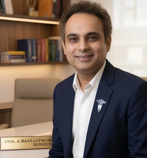
Dr. Sanmit Joshi
Expertise in Oral Lesion Diagnosis and Maxillofacial Surgery
Dr. Sanmit Joshi is an Oral & Maxillofacial Surgeon with an MDS and a Fellowship in Head & Neck Surgical Oncology from Tata Memorial Hospital, Mumbai. His clinical work includes the assessment and surgical management of complex oral and maxillofacial conditions, including lesions that may require further oncological treatment or reconstruction.
He consults at Jaslok Hospital and Research Centre, VED Hospital in Dadar, and Baysmiles Clinic at Kemps Corner, Mumbai. His approach involves clinical evaluation, appropriate investigations, tissue diagnosis, and treatment planning based on the findings.
Years of Post-Fellowship Experience
Years in Oral & Maxillofacial Surgery
Advanced Surgical Training Programs
Specialized Areas of Surgical Expertise
Frequently Asked Questions About Oral & Maxillofacial Pathology Surgery
Does every oral lesion need surgery?
No. Some lesions may resolve after treating the underlying cause, while others can be monitored. A biopsy or surgical procedure may be recommended if the lesion persists, has suspicious features, or requires a confirmed diagnosis.
How long does it take to receive a biopsy report?
The time required varies according to the specimen and the laboratory tests needed. Your treating team can advise when the report is expected and arrange a follow-up consultation to explain the findings.
Can a benign oral lesion return after removal?
Yes. Some lesions can recur depending on their type, extent, and whether they were completely removed. The need for follow-up depends on the final diagnosis and the known recurrence risk.
What is the difference between a biopsy and complete lesion removal?
A biopsy involves collecting tissue for examination, whereas complete removal aims to excise the entire lesion. Some small lesions can be removed during an excisional biopsy, while larger or suspicious lesions may need a separate biopsy before definitive treatment.
Can oral pathology surgery affect my teeth or jaw?
The possibility depends on the lesion’s location and extent. Some procedures involve only the soft tissues, while lesions close to tooth roots or within the jawbone may require more extensive treatment. The expected risks are discussed during surgical planning.
Is oral and maxillofacial pathology surgery painful?
Anaesthesia is used during the procedure to control pain. Some soreness, swelling, or discomfort may occur afterward, depending on the extent of surgery, and can usually be managed with the prescribed aftercare plan.
Will I need a scan before oral pathology surgery?
Not always. Small, superficial lesions may be assessed clinically, while lesions involving the jawbone, deeper tissues, or complex anatomical areas may require dental X-rays, CBCT, CT, or MRI.
What happens if the biopsy confirms oral cancer?
Further assessment is required to determine the extent of the disease and the appropriate treatment. Depending on the findings, care may involve cancer surgery, neck assessment, reconstruction, or other treatments coordinated with the relevant specialist team.
Get an Evaluation for Oral & Maxillofacial Pathology Surgery in Mumbai
Persistent oral lesions, unexplained swellings, and abnormal jaw findings should be assessed to establish the cause and determine the appropriate next step. Book a Consultation with Dr. Sanmit Joshi to discuss your symptoms, investigation findings, and available treatment options.
